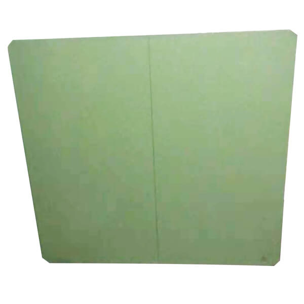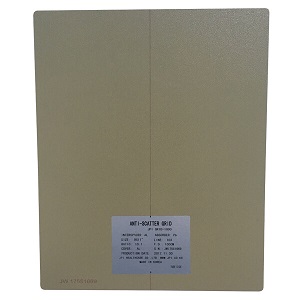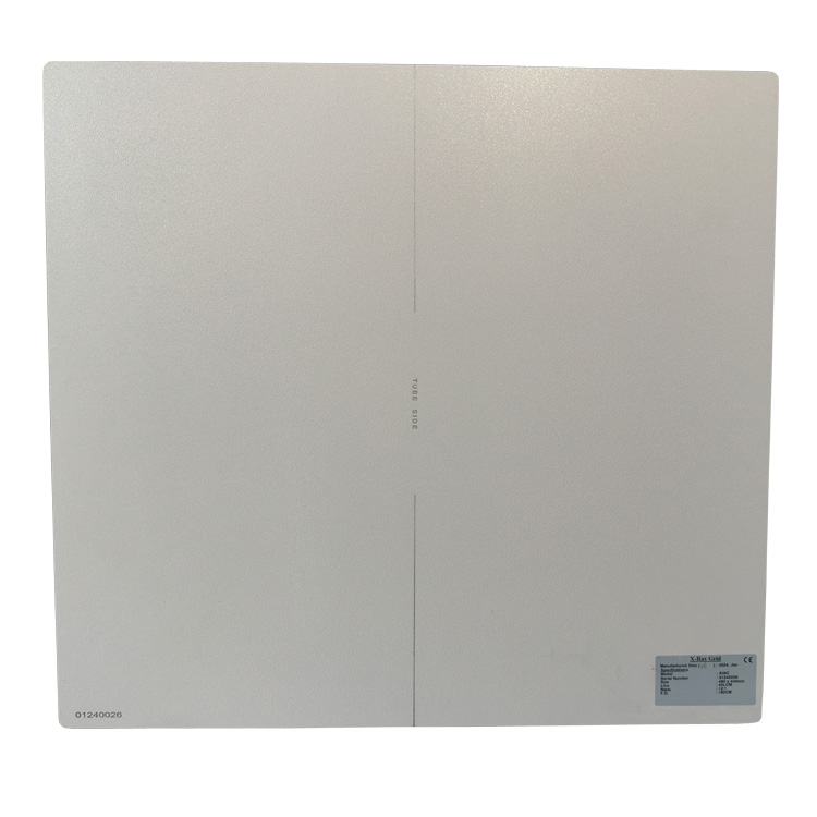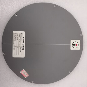
Grids in radiography are placed between the film and the limbs. The centerline of the X-ray tube is aligned with the center of the grid plate, so that the primary X-rays emitted from the X-ray tube are parallel to the grid lead, and part of it passes through the lead. The gap between the strips reaches the film, and the other part of the rays is absorbed on the lead strip. The scattered rays emitted by the subject cannot pass through the lead strip gap because of the angle with the lead strip, so most of it is absorbed, thereby reducing the amount of light on the photo. The amount of scattered rays greatly improves the contrast of the photo and improves the image quality of the photo.
The higher the grids in radiography ratio, the stronger the ability to eliminate scattered rays and the greater the patient’s exposure. The greater the grid density, the greater the dose received by the patient. Therefore, when using grids in radiography, it is not that the higher the grid ratio, the greater the grid density, the better. In daily work, it should be determined according to the size of the frequently used kV. For example, when using high-kV photography, it is best to use grids in radiography of 10:1 or 12:1. When photographing parts with a thickness of more than 14cm, it is generally used. 8:1 or 10:1 grids in radiography will do. The grid with high grid density is generally a fixed grid.
Author:Lillian
Tel: +86 17616362240
Email: newheek1999@outlook.com
Company: Weifang Newheek Electronic Tech Co., Ltd.
Address: E Building of Future Star Scientific Innovation Industrial Zone of No.957 Wolong East Street, Yulong Community, Xincheng Sub-District Office, Weifang Hi-tech Zone, Shandong Province, China